Application Progress on Transmission Electron Microscopy in the Research of Rare Earth Deposits
-
摘要:
透射电子显微镜(简称“透射电镜”)技术具非常高的空间分辨率和多样化的测试功能,可以获取样品非常全面的亚微米-纳米尺度的晶体结构和化学成分信息,是研究地质样品微观组成和结构的强有力工具,近年来越来越多地应用到包括矿床学在内的地球科学研究领域。本文以透射电镜在关键金属稀土矿产资源研究中的应用为例,在简述透射电镜的基本结构、工作原理、功能和制样方法基础上,重点综述了其在揭示碳酸岩型、伟晶岩型、风化壳离子吸附型、磷块岩型和深海沉积物型稀土矿床成矿特征和成因模式研究中的应用进展,包括氟碳铈矿的颗粒附着结晶生长机制,磷灰石和锆石受热液交代导致的矿物溶解、迁移和再沉淀过程,离子吸附型矿床中原生稀土矿物的微观结构和风化产物特征,磷灰石中稀土元素的纳米-原子尺度的赋存状态和替代机制等。提出了透射电镜研究过程中需要注意的事项和应用前景。
Abstract:Transmission electron microscopy (TEM), having a remarkably high spatial resolution and diversified analysis capability, can be used to obtain very comprehensive composition, structure, and crystal chemistry of the samples under submicron-nanometer scale and is a powerful tool for studying the microscopic components and structure of geological samples. It has been applied increasingly in earth science, including mineral deposits. In this paper, TEM is used as an example of studying rare earth deposits. Firstly, we briefly introduce the TEM’s fundamental structure, working principles, function, and sample preparation method. Then, we review the application of TEM in the study of rare earth element (REE) deposits from carbonatite-, pegmatite-, granitoid weathering crust-, and phosphorite- to deep-sea sediment types. The work involves the bastnaesite’s growth mechanism of crystallization of particle attachment, the apatite and zircon’s dissolution, migration, and re-precipitation during hydrothermal metasomatism, the microstructure of primary REEs and the mineral types of weathering products in the granitoid weathering crust deposits, the apatite’s occurrence state and substitution mechanism of trace REEs at the nano to atom scale. Finally, the cautions and application prospects for the TEM in studying geology in the future are summarized.
-

-
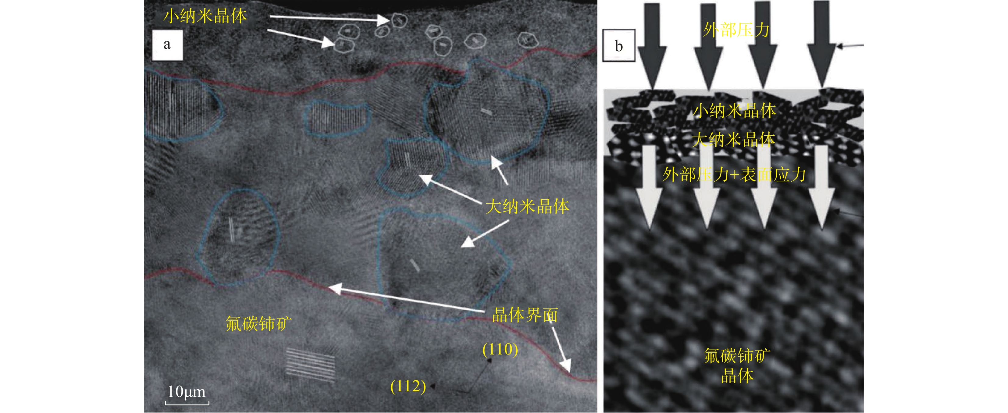
图 2 氟碳铈矿晶体高分辨图和氟碳铈矿的生长模式图(修改自Liu等[39])
Figure 2.
图 3 颗粒附着结晶(CPA)晶体生长机制示意图(修改自de Yoreo等[40])
Figure 3.
图 5 波兰南部Piława Gorna伟晶岩中锆石-磷钇矿蚀变结构照片(修改自Tramm等[48])
Figure 5.
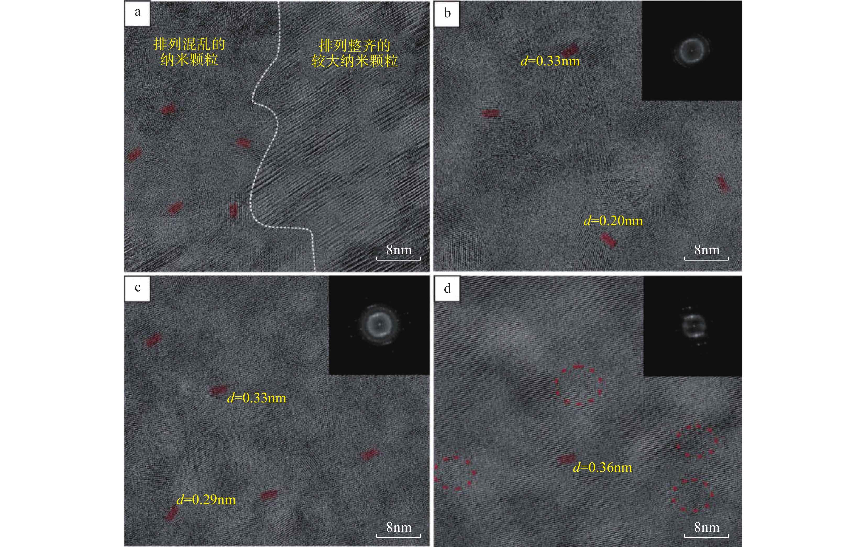
图 6 波兰南部Piława Gorna伟晶岩中磷钇矿透射电镜图像(修改自Tramm等[48])
Figure 6.
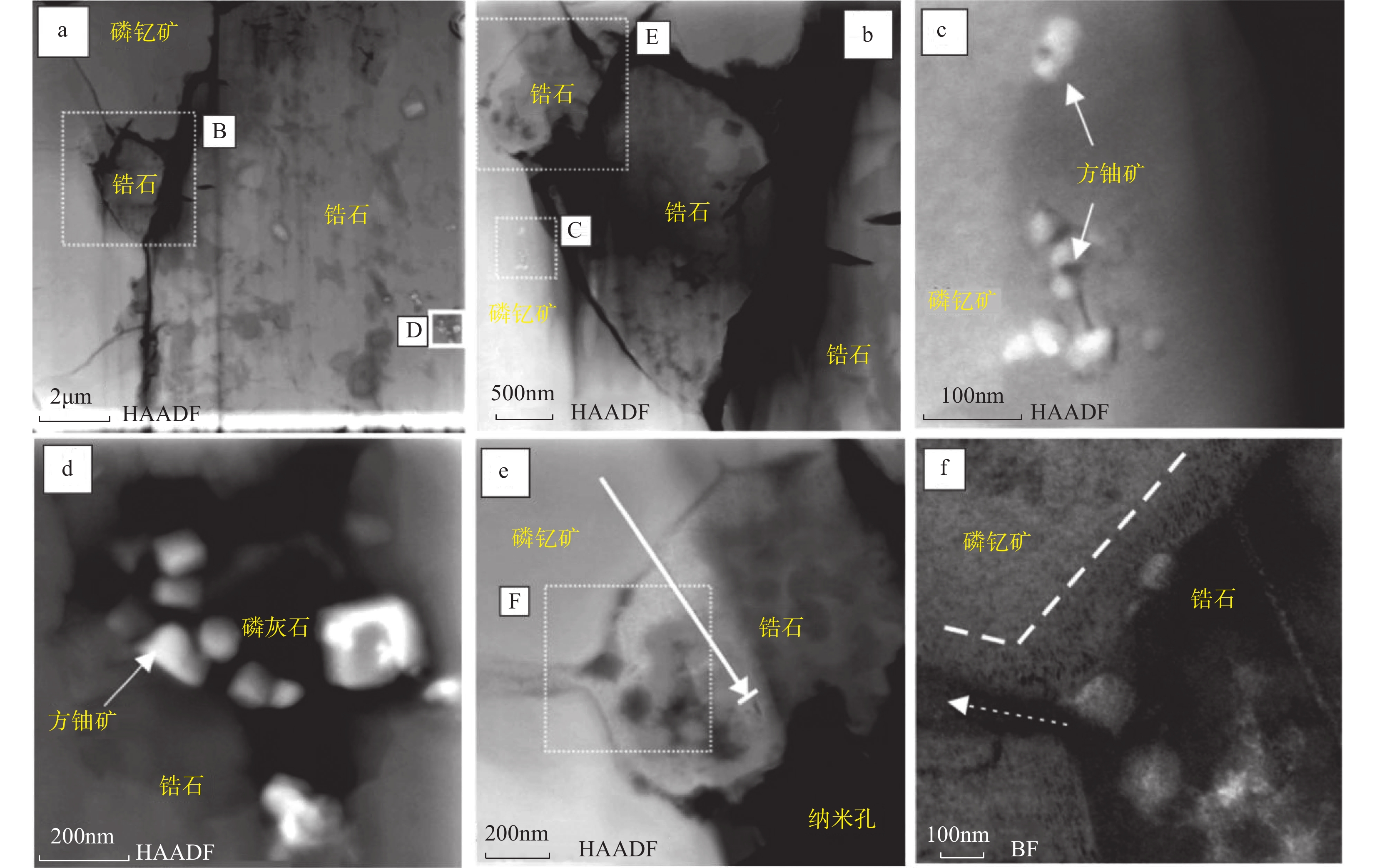
图 7 波兰南部Piława Gorna伟晶岩中锆石-磷钇矿透射电镜图像(修改自Tramm等[48])
Figure 7.
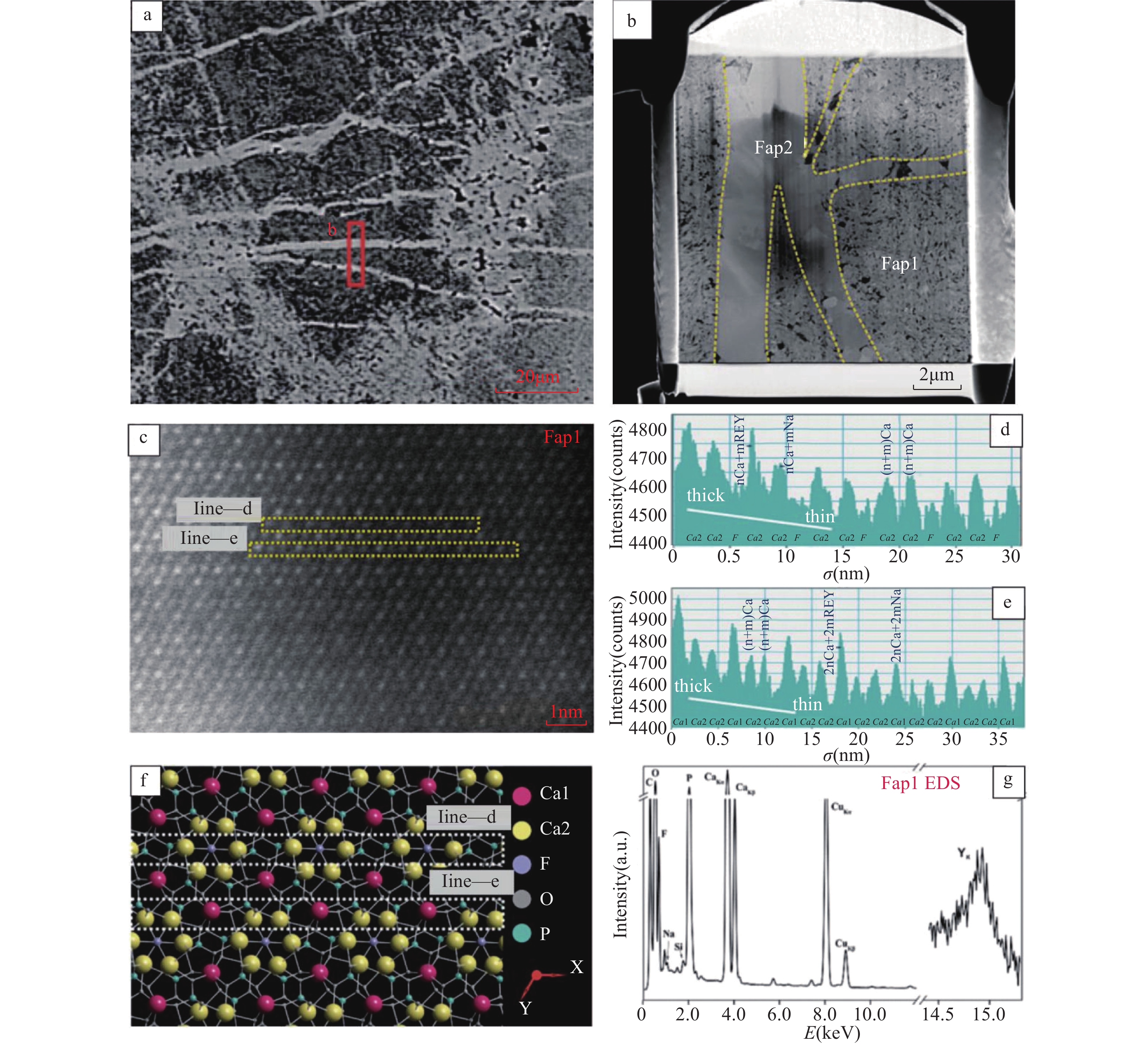
图 8 江西寨背风化壳型稀土矿床中风化花岗岩中稀土矿物的透射电镜图像(修改自Shi等[54])
Figure 8.
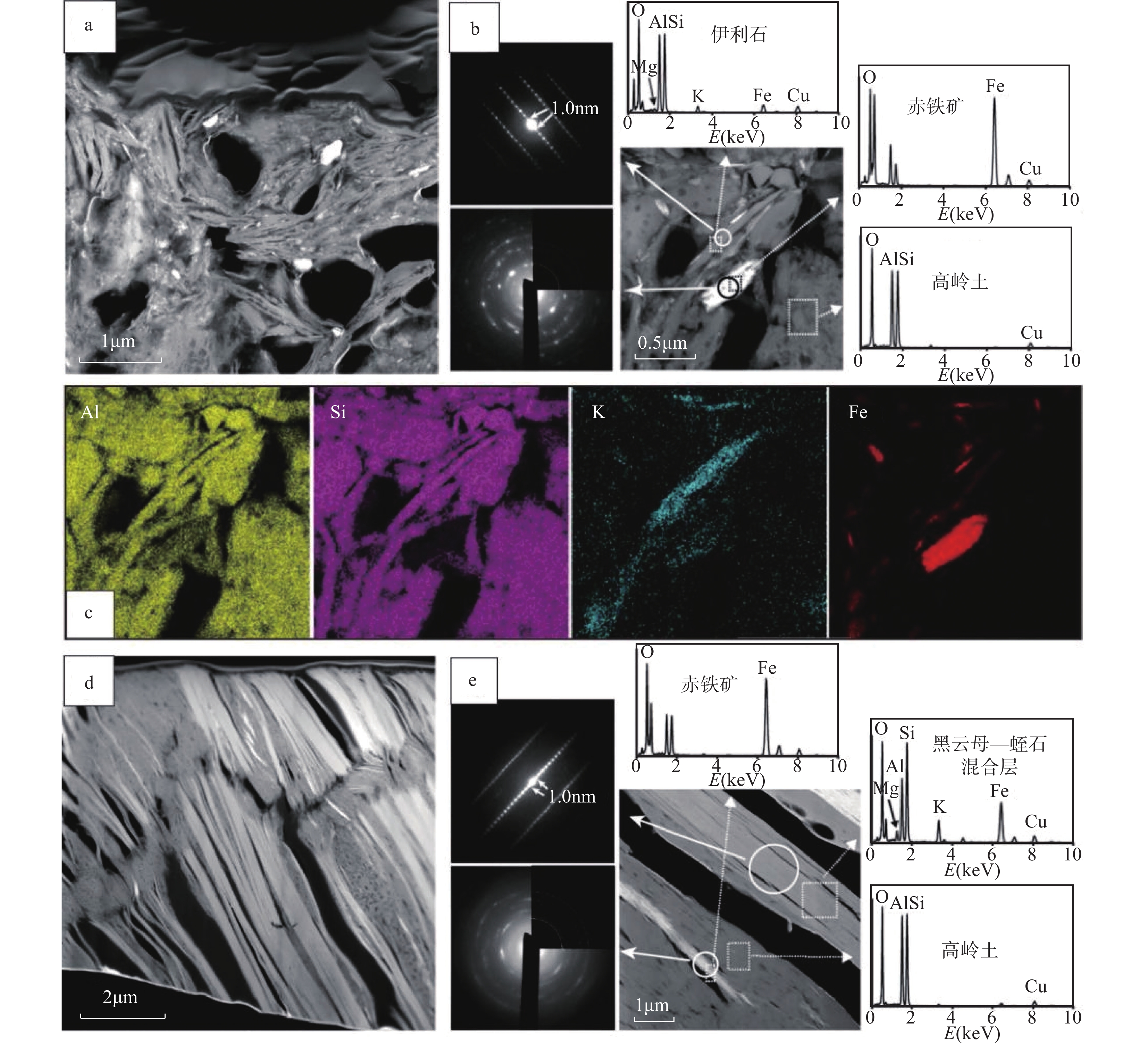
图 9 中国某花岗岩风化壳型稀土矿床中钾长石和黑云母风化产物的透射电镜图片(修改自Mukai等[55])
Figure 9.
图 10 贵州磷块岩中磷灰石的扫描电镜和透射电镜图片(修改自Xing等[61])
Figure 10.
图 11 深海沉积物中羟基磷灰石的透射电镜照片(修改自Liao等[64])
Figure 11.
-
[1] 李金华, 潘永信. 透射电子显微镜在地球科学研究中的应用[J]. 中国科学: 地球科学, 2015, 45(9): 1359−1382. doi: 10.1360/zd2015-45-9-1359
Li J H, Pan Y X. Applications of transmission electron microscopy in the Earth sciences[J]. Scientia Sinica Terrae, 2015, 45(9): 1359−1382. doi: 10.1360/zd2015-45-9-1359
[2] 琚宜文, 孙岩, 万泉, 等. 纳米地质学: 地学领域革命性挑战[J]. 矿物岩石地球化学通报, 2016, 35(1): 1−20, 22−23. doi: 10.3969/j.Issn.1007-2802.2016.01,001
Ju Y W, Sun Y, Wan Q, et al. Nanogeology: A revolutionary challenge in geosciences[J]. Bulletin of Mineralogy, Petrology and Geochemistry, 2016, 35(1): 1−20, 22−23. doi: 10.3969/j.Issn.1007-2802.2016.01,001
[3] 琚宜文, 黄骋, 孙岩, 等. 纳米地球科学: 内涵与意义[J]. 地球科学, 2018, 43(5): 1367−1383. doi: CNKI:SUN:DQKX.0.2018-05-002
Ju Y W, Hang C, Sun Y, et al. Nanogeoscience: Connotation and significance[J]. Earth Science, 2018, 43(5): 1367−1383. doi: CNKI:SUN:DQKX.0.2018-05-002
[4] 唐旭, 李金华. 透射电子显微镜技术新进展及其在地球和行星科学研究中的应用[J]. 地球科学, 2021, 46(4): 1374−1415. doi: 10.3799/dqkx.2020.387
Tang X, Li J H. Transmission electron microscopy: New advances and applications for Earth and Planetary sciences[J]. Earth Science, 2021, 46(4): 1374−1415. doi: 10.3799/dqkx.2020.387
[5] 何宏平, 朱建喜, 陈锰, 等. 矿物结构与矿物物理研究进展综述(2011~2020年)[J]. 矿物岩石地球化学通报, 2020, 39(4): 697−713, 682. doi: 10.19658/j.issn.1007-2802.2020.39.058
Hong H P, Zhu J X, Chen M, et al. Progresses in researches on mineral structure and mineral physics (2011—2020)[J]. Bulletin of Mineralogy, Petrology and Geochemistry, 2020, 39(4): 697−713, 682. doi: 10.19658/j.issn.1007-2802.2020.39.058
[6] Wu X, Meng D, Han Y. α-PbO2-type nanophase of TiO2 from coesite-bearing eclogite in the Dabie Mountains, China[J]. American Mineralogist, 2005, 90(8−9): 1458−1461. doi: 10.2138/am.2005.1901
[7] 孟大维, 吴秀玲, 孙凡, 等. 大别山硬玉石英岩中发现α-PbO2型TiO2超高压相[J]. 地球科学(中国地质大学学报), 2008, 33(5): 706−715. doi: CNKI:SUN:DQKX.0.2008-05-018
Meng D W, Wu X L, Sun F, et al. Identification of α-PbO2-type TiO2 in jadeite quartzite from Shuanghe‚ Dabie Mountains, China[J]. Earth Science—Journal of China University of Geosciences, 2008, 33(5): 706−715. doi: CNKI:SUN:DQKX.0.2008-05-018
[8] Palenik C S, Utsunomiya S, Reich M, et al. Invisible, gold revealed: Direct imaging of gold nanoparticles in a Carlin-type deposit[J]. American Mineralogist, 2004, 89(10): 1359−1366. doi: 10.2138/am-2004-1002
[9] McLeish D F, Williams-Jones A E, Vasyukova O V, et al. Colloidal transport and flocculation are the cause of the hyperenrichment of gold in nature[J]. Proceedings of the National Academy of Sciences, 2021, 118(20): e2100689118. doi: 10.1073/pnas.2100689118
[10] Meng L, Zhu S, Li X, et al. Incorporation mechanism of structurally bound gold in pyrite: Insights from an integrated chemical and atomic-scale microstructural study[J]. American Mineralogist: Journal of Earth and Planetary Materials, 2022, 107(4): 603−613. doi: 10.2138/am-2021-7812
[11] Gamaletsos P N, Godelitsas A, Kasama T, et al. Nano-mineralogy and-geochemistry of high-grade diasporic karst-type bauxite from Parnassos—Ghiona mines, Greece[J]. Ore Geology Reviews, 2017, 84: 228−244. doi: 10.1016/j.oregeorev.2016.11.009
[12] Liu X, Liu R, Chen G, et al. Natural HgS nanoparticles in sulfide minerals from the Hetai goldfield[J]. Environmental Chemistry Letters, 2020, 18(3): 941−947. doi: 10.1007/s10311-020-00978-y
[13] 范宏瑞, 牛贺才, 李晓春, 等. 中国内生稀土矿床类型、成矿规律与资源展望[J]. 科学通报, 2020, 65(33): 3778−3793. doi: 10.1360/TB-2020-0432
Fang H R, Niu H C, Li X C, et al. The types, ore genesis and resource perspective of endogenic REE deposits in China[J]. Chinese Science Bulletin, 2020, 65(33): 3778−3793. doi: 10.1360/TB-2020-0432
[14] 宋文磊, 许成, 王林均, 等. 与碳酸岩碱性杂岩体相关的内生稀土矿床成矿作用研究进展[J]. 北京大学学报(自然科学版), 2013, 49(4): 725−740. doi: 10.13209/j.0479-8023.2013.100
Song W L, Xu C, Wang L J, et al. Review of the metallogenesis of the endogenetic rare earth elements deposits related to carbonatite-alkaline complex[J]. Acta Scientiarum Naturalium Universitatis Pekinensis, 2013, 49(4): 725−740. doi: 10.13209/j.0479-8023.2013.100
[15] Xie Y, Hou Z, Goldfarb R J, et al. Rare earth element deposits in China[M]. United States: Reviews in Economic Geology, 2016: 115−136.
[16] 李斗星. 透射电子显微学的新进展Ⅰ. 透射电子显微镜及相关部件的发展及应用[J]. 电子显微学报, 2004, 23(3): 269−277. doi: 10.3969/j.issn.1000-6281.2004.03.017
Li D X. Progress of transmission electron microscopy Ⅰ. Development of transmission electron microscope and related equipments[J]. Journal of Chinese Electron Microscopy Society, 2004, 23(3): 269−277. doi: 10.3969/j.issn.1000-6281.2004.03.017
[17] 尹美杰, 健男, 张熙, 等. 透射电子显微镜空间分辨率综述[J]. 深圳大学学报(理工版), 2023, 40(1): 1−13. doi: 10.3724/SP.J.1249.2023.01001
Yin M J, Jian N, Zhang X, et al. Review on the spatial resolution of transmission electron microscope[J]. Journal of Shenzhen University Science and Engineering (Science & Engineering), 2023, 40(1): 1−13. doi: 10.3724/SP.J.1249.2023.01001
[18] 贾志宏, 丁立鹏, 陈厚文. 高分辨扫描透射电子显微镜原理及其应用[J]. 物理, 2015, 44(7): 446−452. doi: 10.7693/wl20150704
Jia Z H, Ding L P, Chen H W. The principle and applications of high-resolution scanning electron microscopy[J]. Physics, 2015, 44(7): 446−452. doi: 10.7693/wl20150704
[19] 谷立新, 李金华. 聚焦离子束显微镜技术及其在地球和行星科学研究中的应用[J]. 矿物岩石地球化学通报, 2020, 39(6): 1119−1140, 1065−1066. doi: 10.19658/j.issn.1007-2802.2020.39.102
Gu L X, Li J H. The focused ion beam (FIB) technology and its applications for Earth and Planetary Sciences[J]. Bulletin of Mineralogy, Petrology and Geochemistry, 2020, 39(6): 1119−1140, 1065−1066. doi: 10.19658/j.issn.1007-2802.2020.39.102
[20] 王磊, 曲迪, 姬静远, 等. 基于FIB-SEM制备尖晶石微米颗粒的球差校正透射电镜样品[J]. 电子显微学报, 2021, 40(1): 50−54. doi: 10.3969/j.issn.1000-6281.2021.01.010
Wang L, Qu J, Ji J Y, et al. Preparation of spherical aberration corrected TEM samples of spinel micro-sized particles based on FIB-SEM[J]. Journal of Chinese Electron Microscopy Society, 2021, 40(1): 50−54. doi: 10.3969/j.issn.1000-6281.2021.01.010
[21] 范宏瑞, 谢奕汉, 王凯怡, 等. 碳酸岩流体及其稀土成矿作用[J]. 地学前缘, 2001, 8(4): 289−295. doi: 10.3321/j.issn:1005-2321.2001.04.008
Fan H R, Xie Y H, Wang K Y, et al. Carbonatitic fluids and REE mineralization[J]. Earth Science Frontiers, 2001, 8(4): 289−295. doi: 10.3321/j.issn:1005-2321.2001.04.008
[22] Xie Y, Li Y, Hou Z, et al. A model for carbonatite hosted REE mineralisation—The Mianning—Dechang REE belt, Western Sichuan Province, China[J]. Ore Geology Reviews, 2015, 70: 595−612. doi: 10.1016/j.oregeorev.2014.10.027
[23] Anenburg M, Broom-Fendley S, Chen W. Formation of rare earth deposits in carbonatites[J]. Elements: An International Magazine of Mineralogy, Geochemistry, and Petrology, 2021, 17(5): 327−332. doi: 10.2138/gselements.17.5.327
[24] 尹淑苹, 谢玉玲, 侯增谦, 等. 碳酸岩研究进展[J]. 岩石学报, 2024, 40(3): 1003−1022. doi: 10.18654/1000-0569/2024.03.18
Yin S P, Xie Y L, Hong Z Q, et al. A review of research advances on carbonatite[J]. Acta Petrologica Sinica, 2024, 40(3): 1003−1022. doi: 10.18654/1000-0569/2024.03.18
[25] 许成, 宋文磊, 何晨, 等. 外生稀土矿床的分布、类型和成因概述[J]. 矿物岩石地球化学通报, 2015, 34(2): 234−241. doi: 10.3969/j.issn.1007-2802.2015.02.002
Xu C, Song W L, He C, et al. The overview of the distribution, type and genesis of the exogenetic rare earth elements (REE) deposits[J]. Bulletin of Mineralogy, Petrology and Geochemistry, 2015, 34(2): 234−241. doi: 10.3969/j.issn.1007-2802.2015.02.002
[26] 王登红, 赵芝, 于扬, 等. 离子吸附型稀土资源研究进展、存在问题及今后研究方向[J]. 岩矿测试, 2013, 32(5): 796−802. doi: 10.15898/j.cnki.11-2131/td.2013.05.005
Wang D H, Zhao Z, Yu Y, et al. Progress, problems and research orientation of ion-adsorption type rare earth resources[J]. Rock and Mineral Analysis, 2013, 32(5): 796−802. doi: 10.15898/j.cnki.11-2131/td.2013.05.005
[27] He C, Xu C, Zhao Z, et al. Petrogenesis and mineralization of REE-rich granites in Qingxi and Guanxi, Nanling region, South China[J]. Ore Geology Reviews, 2017, 81: 309−325. doi: 10.1016/j.oregeorev.2016.10.021
[28] Li Y H M, Zhao W W, Zhou M F. Nature of parent rocks, mineralization styles and ore genesis of regolith-hosted REE deposits in South China: An integrated genetic model[J]. Journal of Asian Earth Sciences, 2017, 148: 65−95. doi: 10.1016/j.jseaes.2017.08.004
[29] 付伟, 赵芹, 罗鹏, 等. 中国南方离子吸附型稀土矿床成矿类型及其母岩控矿因素探讨[J]. 地质学报, 2022, 96(11): 3901−3925. doi: 10.19762/j.cnki.dizhixuebao.2022233
Fu W, Zhao Q, Luo P, et al. Mineralization diversity of ion adsorption type REE deposit in Southern China and the critical influence of parent rocks[J]. Acta Geologica Sinica, 2022, 96(11): 3901−3925. doi: 10.19762/j.cnki.dizhixuebao.2022233
[30] 张杰, 孙传敏, 龚美菱, 等. 贵州织金含稀土生物屑磷块岩稀土元素赋存状态研究[J]. 稀土, 2007(1): 75−79. doi: 10.16533/j.cnki.15-1099/tf.2007.01.021
Zhang J, Sun C M, Gong M L, et al. Geochemical characteristics and occurrence states of the REE elements of the phosphorite in Xinhua, Zhijin, Guizhou[J]. Chinese Rare Earths, 2007(1): 75−79. doi: 10.16533/j.cnki.15-1099/tf.2007.01.021
[31] 陈吉艳, 杨瑞东, 张杰. 贵州织金含稀土磷矿床稀土元素赋存状态研究[J]. 矿物学报, 2010, 30(1): 123−129. doi: 10.16461/j.cnki.1000-4734.2010.01.012
Chen J Y, Yang R D, Zhang J. Mode of occurrence of rare earth elements in posphorite in Zhijin County, Guizhou Province, China[J]. Acta Mineralogica Sinica, 2010, 30(1): 123−129. doi: 10.16461/j.cnki.1000-4734.2010.01.012
[32] 谢宏, 朱立军. 贵州早寒武世早期磷块岩稀土元素赋存状态及分布规律研究[J]. 中国稀土学报, 2012, 30(5): 620−627. doi: CNKI:SUN:XTXB.0.2012-05-018
Xie H, Zhu L J. Existing state and distribution regularity of rare earth elements from early Cambrian phosphorite in Guizhou[J]. Journal of the Chinese Society of Rare Earths, 2012, 30(5): 620−627. doi: CNKI:SUN:XTXB.0.2012-05-018
[33] Kon Y, Hoshino M, Sanematsu K, et al. Geochemical characteristics of apatite in heavy REE-rich deep-sea mud from Minami—Torishima area, Southeastern Japan[J]. Resource Geology, 2014, 64(1): 47−57. doi: 10.1111/rge.12026
[34] 石学法, 符亚洲, 李兵, 等. 我国深海矿产研究: 进展与发现(2011—2020)[J]. 矿物岩石地球化学通报, 2021, 40(2): 305−318, 517. doi: 10.19658/j.issn.1007-2802.2021.40.022
Shi F X, Fu Y Z, Li B, et al. Research on deep-sea minerals in China: Progress and discovery (2011-2020)[J]. Bulletin of Mineralogy, Petrology and Geochemistry, 2021, 40(2): 305−318, 517. doi: 10.19658/j.issn.1007-2802.2021.40.022
[35] 樊文枭, 周军明, 张欢, 等. 富稀土深海沉积物中稀土元素载体矿物的研究进展与展望[J]. 矿物学报, 2023, 43(2): 145−156. doi: 10.16461/j.cnki.1000-4734.2023.43.012
Fan W X, Zhou J M, Zhang H, et al. Research progresses and prospects on carrier minerals of rare earth elements (REY) in deep-sea REY-rich sediments[J]. Acta Mineralogica Sinica, 2023, 43(2): 145−156. doi: 10.16461/j.cnki.1000-4734.2023.43.012
[36] Castor S B. Rare earth deposits of North America[J]. Resource Geology, 2008, 58(4): 337−347. doi: 10.1111/j.1751-3928.2008.00068.x
[37] Liu Y, Chakhmouradian A R, Hou Z, et al. Development of REE mineralization in the giant Maoniuping deposit (Sichuan, China): Insights from mineralogy, fluid inclusions, and trace-element geochemistry[J]. Mineralium Deposita, 2018, 54: 701−718. doi: 10.1007/s00126-018-0836-y
[38] Song W, Xu C, Smith M P, et al. Genesis of the world’s largest rare earth element deposit, Bayan Obo, China: Protracted mineralization evolution over 1b.y.[J]. Geology, 2018, 46(4): 323−326. doi: 10.1130/G39801.1
[39] Liu R, Bo B, Tao D, et al. Bastnäsite nanoparticles in carbonatite-syenite-hosted REE deposit: Implication for La and Ce migration and bastnäsite growth[J]. Chemosphere, 2021, 271: 129831. doi: 10.1016/j.chemosphere.2021.129831
[40] de Yoreo J J, Gilbert P U P A, Sommerdijk N A J M, et al. Crystallization by particle attachment in synthetic, biogenic, and geologic environments[J]. Science, 2015, 349(6247): aaa6760. doi: 10.1126/science.aaa6760
[41] Wall F, Niku-Paavola V N, Storey C, et al. Xenotime-(Y) from carbonatite dykes at Lofdal, Namibia: Unusually low LREE: HREE ratio in carbonatite, and the first dating of xenotime overgrowths on zircon[J]. The Canadian Mineralogist, 2008, 46(4): 861−877. doi: 10.3749/canmin.46.4.861
[42] Chakhmouradian A R, Wall F. Rare earth elements: Minerals, mines, magnets (and more)[J]. Elements, 2012, 8(5): 333−340. doi: 10.2113/gselements.8.5.333
[43] Trofanenko J, Williams-Jones A E, Simandl G J, et al. The nature and origin of the REE mineralization in the Wicheeda carbonatite, British Columbia, Canada[J]. Economic Geology, 2016, 111(1): 199−223. doi: 10.2113/econgeo.111.1.199
[44] 许丹妮, 宋文磊, 杨金昆, 等. 内生碳酸岩型稀土矿床中磷酸盐作用研究综述[J]. 地质论评, 2024, 70(3): 1097−1112. doi: 10.5382/SP.22.12
Xu D N, Song W L, Yang J K, et al. A review on the role of phosphate in endogenetic carbonatite-hosted rare-earth deposits[J]. Geological Review, 2024, 70(3): 1097−1112. doi: 10.5382/SP.22.12
[45] Harlov D E, Andersson U B, Förster H J, et al. Apatite–monazite relations in the Kiirunavaara magnetite–apatite ore, Northern Sweden[J]. Chemical Geology, 2002, 191(1−3): 47−72. doi: 10.1016/S0009-2541(02)00148-1
[46] Harlov D E, Wirth R, Förster H J. An experimental study of dissolution–reprecipitation in fluorapatite: Fluid infiltration and the formation of monazite[J]. Contributions to Mineralogy and Petrology, 2005, 150: 268−286.
[47] Harlov D E. Apatite: A fingerprint for metasomatic processes[J]. Elements, 2015, 11(3): 171−176. doi: 10.2113/gselements.11.3.171
[48] Tramm F, Wirth R, Budzyń B, et al. LA-ICP-MS and TEM constraints on the magmatic and post-magmatic processes recorded by the zircon-xenotime intergrowth in pegmatite (Piława Górna, Góry Sowie Block, SW Poland)[J]. Lithos, 2021, 404: 106480. doi: 10.1016/j.lithos.2021.106480
[49] Putnis A. Mineral replacement reactions: From macroscopic observations to microscopic mechanisms[J]. Mineralogical Magazine, 2002, 66(5): 689−708. doi: 10.1180/0026461026650056
[50] Bao Z, Zhao Z. Geochemistry of mineralization with exchangeable REY in the weathering crusts of granitic rocks in South China[J]. Ore Geology Reviews, 2008, 33(3-4): 519−535. doi: 10.1016/j.oregeorev.2007.03.005
[51] Xu C, Kynický J, Smith M P, et al. Origin of heavy rare earth mineralization in South China[J]. Nature Communications, 2017, 8(1): 14598. doi: 10.1038/ncomms14598
[52] Ishihara S, Hua R, Hoshino M, et al. REE abundance and REE minerals in granitic rocks in the Nanling range, Jiangxi Province, Southern China, and generation of the REE-rich weathered crust deposits[J]. Resource Geology, 2008, 58(4): 355−372. doi: 10.1111/j.1751-3928.2008.00070.x
[53] Wang M, Li M Y, Zhou M F, et al. Enrichment of rare earth elements during the weathering of alkaline igneous systems: Insights from the Puxiong regolith-hosted rare earth element deposit, SW China[J]. Economic Geology, 2024, 119(1): 161−187. doi: 10.5382/econgeo.5024
[54] Shi A, Xu C, Chakhmouradian A R, et al. Nanostructure reveals REE mineral crystallization mechanisms in granites from a heavy REE deposit, South China[J]. American Mineralogist, 2022, 107(12): 2261−2271. doi: 10.2138/am-2022-8309
[55] Mukai H, Kon Y, Sanematsu K, et al. Microscopic analyses of weathered granite in ion-adsorption rare earth deposit of Jiangxi Province, China[J]. Scientific Reports, 2020, 10(1): 20194. doi: 10.1038/s41598-020-76981-8
[56] Emsbo P, McLaughlin P I, Breit G N, et al. Rare earth elements in sedimentary phosphate deposits: Solution to the global REE crisis?[J]. Gondwana Research, 2015, 27(2): 776−785. doi: 10.1016/j.gr.2014.10.008
[57] Zhang H, Fan H, Wen H, et al. Controls of REY enrichment in the early Cambrian phosphorites[J]. Geochimica et Cosmochimica Acta, 2022, 324: 117−139. doi: 10.1016/j.gca.2022.03.003
[58] Xing J, Jiang Y, Xian H, et al. Hydrothermal activity during the formation of REY-rich phosphorites in the early Cambrian Gezhongwu Formation, Zhijin, South China: A micro-and nano-scale mineralogical study[J]. Ore Geology Reviews, 2021, 136: 104224. doi: 10.1016/j.oregeorev.2021.104224
[59] Zhang Z, Jiang Y, Niu H, et al. Enrichment of rare earth elements in the early Cambrian Zhijin phosphorite deposit, SW China: Evidence from francolite micro-petrography and geochemistry[J]. Ore Geology Reviews, 2021, 138: 104342. doi: 10.1016/j.oregeorev.2021.104342
[60] Kato Y, Fujinaga K, Nakamura K, et al. Deep-sea mud in the Pacific Ocean as a potential resource for rare-earth elements[J]. Nature Geoscience, 2011, 4(8): 535−539. doi: 10.1038/NGEO1185
[61] Xing J, Jiang Y, Xian H, et al. Hydrothermal alteration and the remobilization of rare earth elements during reprecipitation of nano-scale apatite in phosphorites[J]. Lithos, 2023, 444: 107113. doi: 10.1016/j.lithos.2023.107113
[62] 郝龙龙, 覃丽禄. 高质量扫描透射电子显微镜高角环形暗场像的实验关键点[J]. 实验科学与技术, 2020, 18(3): 7−10. doi: 10.12179/1672-4550.20190123
Hao L L, Qin L L. The experimental key points in acquiring high quality HAADF-STEM images[J]. Experiment Science and Technology, 2020, 18(3): 7−10. doi: 10.12179/1672-4550.20190123
[63] Hughes J M, Cameron M, Crowley K D. Structural variations in natural F, OH, and Cl apatites[J]. American Mineralogist, 1989, 74(7−8): 870−876. doi: 10.1016/0024-4937(89)90008-X
[64] Liao J, Sun X, Li D, et al. New insights into nanostructure and geochemistry of bioapatite in REE-rich deep-sea sediments: LA-ICP-MS, TEM, and Z-contrast imaging studies[J]. Chemical Geology, 2019, 512: 58−68. doi: 10.1016/j.chemgeo.2019.02.039
[65] Egerton R F, Li P, Malac M. Radiation damage in the TEM and SEM[J]. Micron, 2004, 35(6): 399−409. doi: 10.1016/j.micron.2004.02.003
[66] Egerton R F. Electron energy-loss spectroscopy in the TEM[J]. Reports on Progress in Physics, 2008, 72(1): 016502. doi: 10.1088/0034-4885/72/1/016502
[67] 陈佳妮. 矿物微区分析中透射电镜测试技术的应用[J]. 高校地质学报, 2021, 27(3): 356−365. doi: 10.16108/j.issn1006-7493.2021037
Chen J N. Application of transmission electron micro-scopy in microarea analysis in mineral[J]. Geological Journal of China Universities, 2021, 27(3): 356−365. doi: 10.16108/j.issn1006-7493.2021037
-


 下载:
下载:
