X-ray CT scanning technique and its application to the Core 01 in the northern South China Sea for sedimentary environment reconstruction
-
摘要:
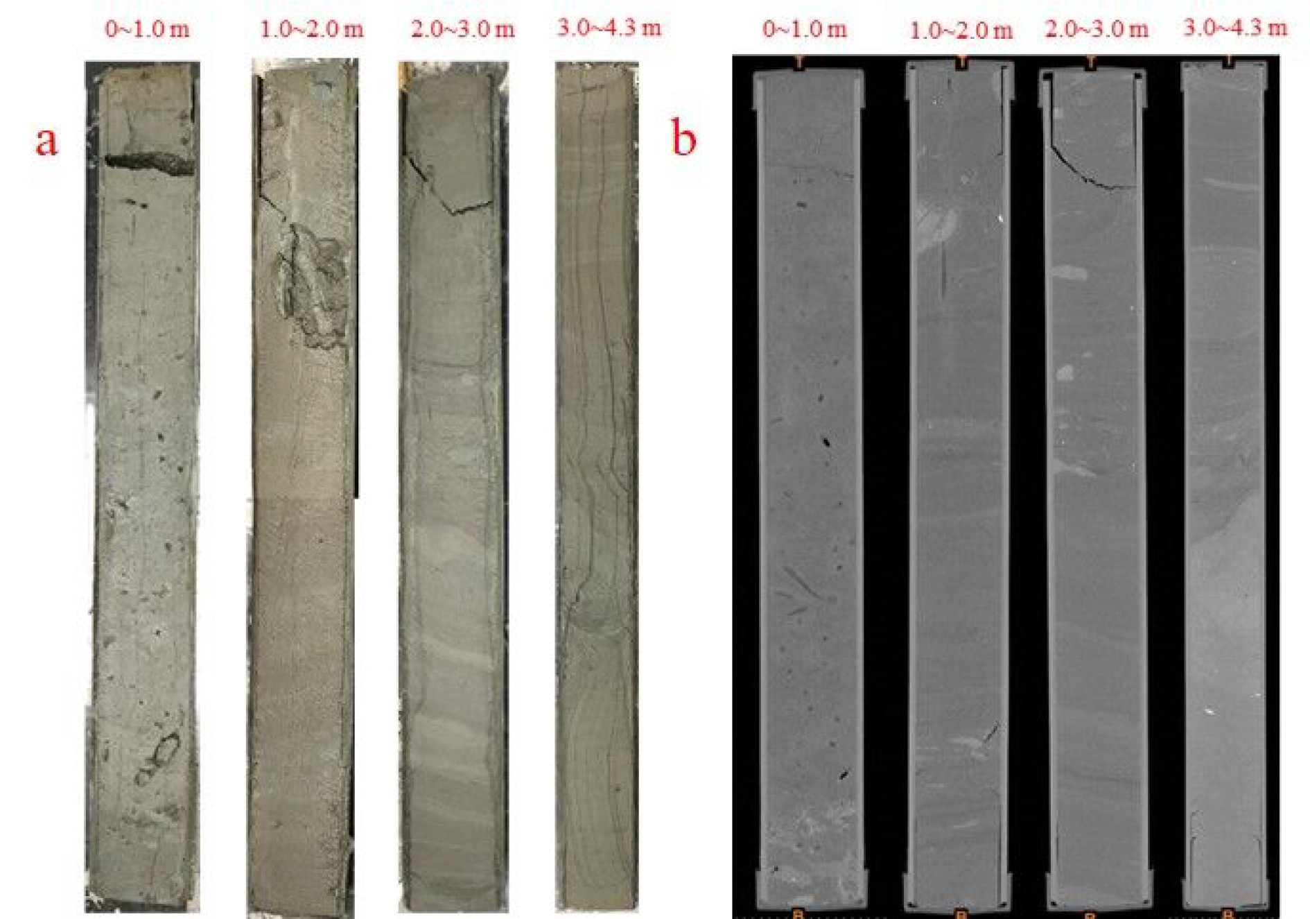
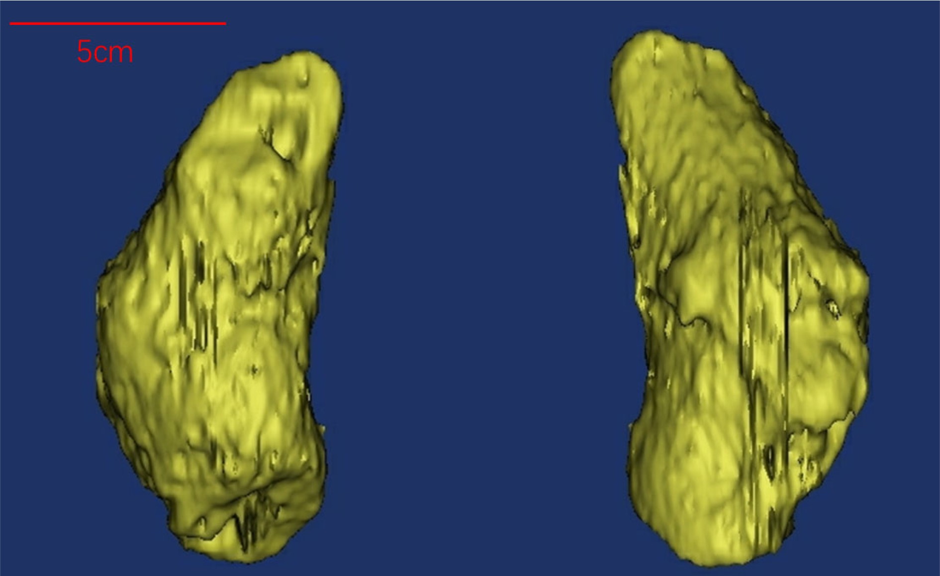
随着技术的不断进步,X射线CT扫描与三维重建技术在地质学研究中的应用越来越广泛,但在深海岩心的分析中应用依然有限。对南海北部陆坡岩心柱Core 01进行全样岩心X射线CT扫描,将扫描图像导入三维建模软件Mimics,进行岩心内部结构的三维重建,恢复了该岩心中0~1.0 m的孔隙结构及4.2 m处的生物化石壳体的外部形态。重建结果表明X射线CT扫描在深海岩心的研究中具有可行性。此外,结合浮游有孔虫AMS 14C测年、底栖有孔虫氧碳同位素结果以及此站位的地质背景,推断该岩心中孔隙的形成可能与该区天然气水合物的释放有关。
Abstract:X-ray CT scanning and three-dimension reconstruction techniques are widely used in geological research nowadays. However, their application to deep sea sediment cores remains rare. In this study, a deep-sea core labeled Core 01 taken from the northern South China Sea was subjected to a full core X-ray CT scan. The images were imputed into Mimics, a three-dimensional modeling software, to carry out 3D reconstruction of the internal structures of the core, which includes the pore structures at 0~1.0 m and the external shape of fossils at 4.2 m. The results confirmed that X-ray CT scan is feasible for study of deep sea sediment cores. In addition, in combination with the AMS 14C dating data of planktonic foraminifera, the oxygen carbon stable isotope results of benthic foraminifera, and the geological background of this region, it is inferred that the formation of pores in the sediment core may be related to the release of natural gas hydrates.
-
Key words:
- X-ray /
- CT scan /
- three-dimension reconstruction /
- sediment core
-

-
[1] 陈世杰, 赵淑萍, 马巍, 等. 利用CT扫描技术进行冻土研究的现状和展望[J]. 冰川冻土, 2013, 35(1):193-200 doi: 10.7522/j.issn.1000-0240.2013.0023
CHEN Shijie, ZHAO Shuping, MA Wei, et al. Studying frozen soil with CT technology: Present studies and prospects [J]. Journal of Glaciology and Geocryology, 2013, 35(1): 193-200. doi: 10.7522/j.issn.1000-0240.2013.0023
[2] 李承峰, 胡高伟, 刘昌岭, 等. X射线计算机断层扫描在天然气水合物研究中的应用[J]. 热带海洋学报, 2012, 31(5):93-99 doi: 10.3969/j.issn.1009-5470.2012.05.014
LI Chengfeng, HU Gaowei, LIU Changling, et al. Application of X-Ray computed tomography in natural gas hydrate research [J]. Journal of Tropical Oceanography, 2012, 31(5): 93-99. doi: 10.3969/j.issn.1009-5470.2012.05.014
[3] 高剑. 三维重建应用系统研究[D]. 山东大学博士学位论文, 2009.
GAO Jian. Research on 3D reconstruction application system[D]. Doctor Dissertation of Shandong University, 2009.
[4] Conroy G C, Vannier M W. Noninvasive three-dimensional computer imaging of Matrix-Filled fossil skulls by high-resolution computed tomography [J]. Science, 1984, 226(4673): 456-458. doi: 10.1126/science.226.4673.456
[5] Zollikofer C P E, de León M S P, Vandermeersch B, et al. Evidence for interpersonal violence in the St. Césaire Neanderthal [J]. Proceedings of the National Academy of Sciences of the United States of America, 2002, 99(9): 6444-6448. doi: 10.1073/pnas.082111899
[6] 牛永斌, 钟建华, 胡斌. 小尺度地质体三维建模研究——以遗迹化石Chondrites和岩心三维建模为例[J]. 古地理学报, 2008, 10(2):207-214 doi: 10.7605/gdlxb.2008.02.011
NIU Yongbin, ZHONG Jianhua, HU Bin. Research of 3D Modeling on small-scale geologic body - taking 3D modeling on trace fossil Chondrites and drilling core as an example [J]. Journal of Palaeogeography, 2008, 10(2): 207-214. doi: 10.7605/gdlxb.2008.02.011
[7] 星耀武, 刘裕生, 苏涛, 等. X-射线CT扫描技术在中新世松属球果化石研究中的应用[J]. 古生物学报, 2010, 49(1):133-137
XING Yaowu, LIU Yusheng, SU Tao, et al. Application of the X-ray CT scanning technique on a late Miocene pine cone from Yunnan, China [J]. Acta Palaeontologica Sinica, 2010, 49(1): 133-137.
[8] 丁奕, 时敏敏, 刘祎楠. 遗迹化石三维重建研究新进展[J]. 地层学杂志, 2016, 40(4):401-410
DING Yi, SHI Minmin, LIU Yinan. New advances in the three-demensional reconstruction of trace fossils [J]. Journal of Stratigraphy, 2016, 40(4): 401-410.
[9] 潘汝江, 何翔, 肖维民, 等. CT扫描技术在岩心三维重建中的应用[J]. CT理论与应用研究, 2018, 27(3):349-356
Pan Rujing, He Xiang, XIAO Weimin, et al. Application of CT scanning technique in core 3D reconstruction [J]. CT Theory and Applications, 2018, 27(3): 349-356.
[10] 徐宗恒, 徐则民, 李凌旭. 基于CT扫描的斜坡非饱和带土体大孔隙定量化研究和三维重建[J]. 水土保持通报, 2015, 35(1):133-138
XU Zongheng, XU Zemin, LI Lingxu. Soil macropores quantification study and 3D reconstruction in Vadose Zones of hillslope based on X-ray computed tomography [J]. Bulletin of Soil and Water Conservation, 2015, 35(1): 133-138.
[11] 王刚, 沈俊男, 褚翔宇, 等. 基于CT三维重建的高阶煤孔裂隙结构综合表征和分析[J]. 煤炭学报, 2017, 42(8):2074-2080
WANG Gang, SHEN Junnan, CHU Xiangyu, et al. Characterization and analysis of pores and fissures of high-rank coal based on CT three-dimensional reconstruction [J]. Journal of China Coal Society, 2017, 42(8): 2074-2080.
[12] Jin S, Takeya S, Hayashi J, et al. Structure analyses of artificial methane hydrate sediments by microfocus X-ray computed tomography [J]. Japanese Journal of Applied Physics, 2004, 43(8R): 5673-5675.
[13] 胡高伟, 李承峰, 业渝光, 等. 沉积物孔隙空间天然气水合物微观分布观测[J]. 地球物理学报, 2014, 57(5):1675-1682 doi: 10.6038/cjg20140530
HU Gaowei, LI Chengfeng, YE Yuguang, et al. Observation of gas hydrate distribution in sediment pore space [J]. Chinese Journal of Geophysics, 2014, 57(5): 1675-1682. doi: 10.6038/cjg20140530
[14] 李承峰, 胡高伟, 张巍, 等. 有孔虫对南海神狐海域细粒沉积层中天然气水合物形成及赋存特征的影响[J]. 中国科学: 地球科学, 2016, 59(11):2223-2230 doi: 10.1007/s11430-016-5005-3
LI Chengfeng, HU Gaowei, ZHANG Wei, et al. Influence of foraminifera on formation and occurrence characteristics of natural gas hydrates in fine-grained sediments from Shenhu area, South China Sea [J]. Science China Earth Sciences, 2016, 59(11): 2223-2230. doi: 10.1007/s11430-016-5005-3
[15] Ashi J. Computed tomography scan image analysis of sediments[C]//Proceeding of the Ocean Drilling Program. Scientific Results. College Station, TX: Ocean Drilling Program, 1997, 156: 151-159.
[16] Tanaka A, Nakano T. Data report: three-dimensional observation and quantification of internal structure of sediment core from Challenger Mound area in the Porcupine Seabight off western Ireland using a medical X-ray CT[C]//Proceeding of IODP, 307. Washington, DC: Integrated Ocean Drilling Program Management International, Inc., 2009.
[17] Orsi T H, Edwards C M, Anderson A L. X-ray computed tomography: a nondestructive method for quantitative analysis of sediment cores [J]. Journal of Sedimentary Research, 1994, 64: 690-693. doi: 10.1306/D4267E74-2B26-11D7-8648000102C1865D
[18] Tanaka A, Nakano T, Ikehara K. X-ray computerized tomography analysis and density estimation using a sediment core from the Challenger Mound area in the Porcupine Seabight, off Western Ireland [J]. Earth, Planets and Space, 2011, 63(2): 103-110. doi: 10.5047/eps.2010.12.006
[19] 王宏语, 孙春岩, 张洪波, 等. 西沙海槽潜在天然气水合物成因及形成地质模式[J]. 海洋地质与第四纪地质, 2005, 25(4):85-91
WANG Hongyu, SUN Chunyan, ZHANG Hongbo, et al. Origin and genetic model of potential gas hydrates in Xisha trough, South China Sea [J]. Marine Geology and Quaternary Geology, 2005, 25(4): 85-91.
[20] 尹希杰, 周怀阳, 杨群慧, 等. 南海北部甲烷渗漏活动存在的证据: 近底层海水甲烷高浓度异常[J]. 海洋学报, 2008, 30(6):69-75
YIN Xijie, ZHOU Huaiyang, YANG Qunhui, et al. The evidence for the existence of methane seepages in the northern South China Sea: abnormal high methane concentration in bottom waters [J]. Acta Oceanologica Sinica, 2008, 30(6): 69-75.
[21] 陈忠, 黄奇瑜, 颜文, 等. 南海西沙海槽的碳酸盐结壳及其对甲烷冷泉活动的指示意义[J]. 热带海洋学报, 2007, 26(2):26-33 doi: 10.3969/j.issn.1009-5470.2007.02.005
CHEN Zhong, HUANG Qiyu, YAN Wen, et al. Authigenic carbonates as evidence for seeping fluids in Xisha trough of South China Sea [J]. Journal of Tropical Oceanography, 2007, 26(2): 26-33. doi: 10.3969/j.issn.1009-5470.2007.02.005
[22] Yang K H, Chu F Y, Zhu Z M, et al. Formation of methane-derived carbonates during the last glacial period on the northern slope of the South China Sea [J]. Journal of Asian Earth Sciences, 2018, 168: 173-185. doi: 10.1016/j.jseaes.2018.01.022
[23] Mackensen A, Wollenburg J, Licari L. Low δ13C in tests of live epibenthic and endobenthic foraminifera at a site of active methane seepage [J]. Paleoceanography, 2006, 21: PA2022.
[24] Martin R A, Nesbitt E A, Campbell K A. The effects of anaerobic methane oxidation on benthic foraminiferal assemblages and stable isotopes on the Hikurangi Margin of eastern New Zealand [J]. Marine Geology, 2010, 272(1-4): 270-284. doi: 10.1016/j.margeo.2009.03.024
[25] Cheng X R, Huang B Q, Jian Z M, et al. Foraminiferal isotopic evidence for monsoonal activity in the South China Sea: a present-LGM comparison [J]. Marine Micropaleontology, 2005, 54(1-2): 125-139. doi: 10.1016/j.marmicro.2004.09.007
[26] Shackleton N J, Hall M A, Pate D. Pliocene stable isotope stratigraphy of Site 846[C]//Proceeding of the Ocean Drilling Program, Scientific Results. College Station, TX: Ocean Drilling Program, 1995, 138: 337-355.
[27] Hill T M, Kennett J P, Valentine D L. Isotopic evidence for the incorporation of methane-derived carbon into foraminifera from modern methane seeps, Hydrate Ridge, Northeast Pacific [J]. Geochimica et Cosmochimica Acta, 2004, 68(22): 4619-4627. doi: 10.1016/j.gca.2004.07.012
[28] Ketcham R A, Carlson W D. Acquisition, optimization and interpretation of X-ray computed tomographic imagery: applications to the geosciences [J]. Computers and Geosciences, 2001, 27(4): 381-400. doi: 10.1016/S0098-3004(00)00116-3
[29] 殷宗军, 黎刚, 朱茂炎. 两种微体化石三维无损成像技术的对比[J]. 微体古生物学报, 2014, 31(4):440-452
YIN Zongjun, LI Gang, ZHU Maoyan. Three dimensional nondestructive imaging techniques for the microfossils: a comparison [J]. Acta Micropalaeontologica Sinica, 2014, 31(4): 440-452.
-


 下载:
下载:


